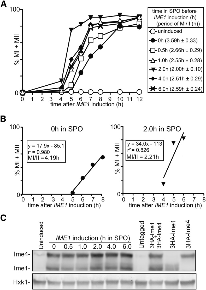
Figure 1.
Synchronous sporulation requires specific timing of IME1 and IME4 induction. (A) Kinetics of meiotic divisions in diploid cells harboring CUP1 promoter fusions with IME1 and IME4 (pCUP-IME1/pCUP-IME4) (FW1810). Cells were grown overnight in rich medium (YPD), diluted to presporulation medium (BYTA), and grown for another 16 hr. Subsequently cells were transferred to sporulation medium (SPO), and IME1 and IME4 were induced at 0, 0.5, 1, 2, 4, and 6 hr in SPO. Samples were collected at 4 hr after induction up to 12 hr with a 1-hr interval, fixed in ethanol, nuclei were stained with DAPI, and DAPI masses were counted. Cells that harbored two, three, or four DAPI masses were classified as cells undergoing meiosis I or meiosis II (% MI + MII, y-axis). For each time point, at least 200 cells were counted. The time after IME1/IME4 induction is plotted on the x-axis. From each time course experiment, we also computed the time or period taken for 75% of the cells to complete meiotic divisions (see Materials and Methods for details). This number is displayed in brackets next to the legend, and represents the mean number of hours from three independent experiments followed by the SEM. (B) Graph to illustrate how we determined the time or period taken to complete meiotic divisions when IME1 and IME4 were induced at 0 or 2 hr after shifting cells to SPO medium as described in (A). A linear trend line was fitted from the first time point where meiotic divisions were detected, to the time point where 75% or more of the cells completed meiotic divisions. From the function, we calculated the period or time taken for 75% of the cells to complete meiotic divisions (MI/II). (C) Western blot showing Ime1 and Ime4 protein levels in cells described in (A). Samples were taken at 2 hr after inducing IME1 and IME4. Ime1 and Ime4 levels were detected by anti-hemagglutinin (HA) antibodies. We also measured Ime1 and Ime4 in an untagged control (FW1511), and in cells that contain HA-tagged IME1 (FW2444), or IME4 (FW2480) alone. To control for loading, Hxk1 levels were also determined.

