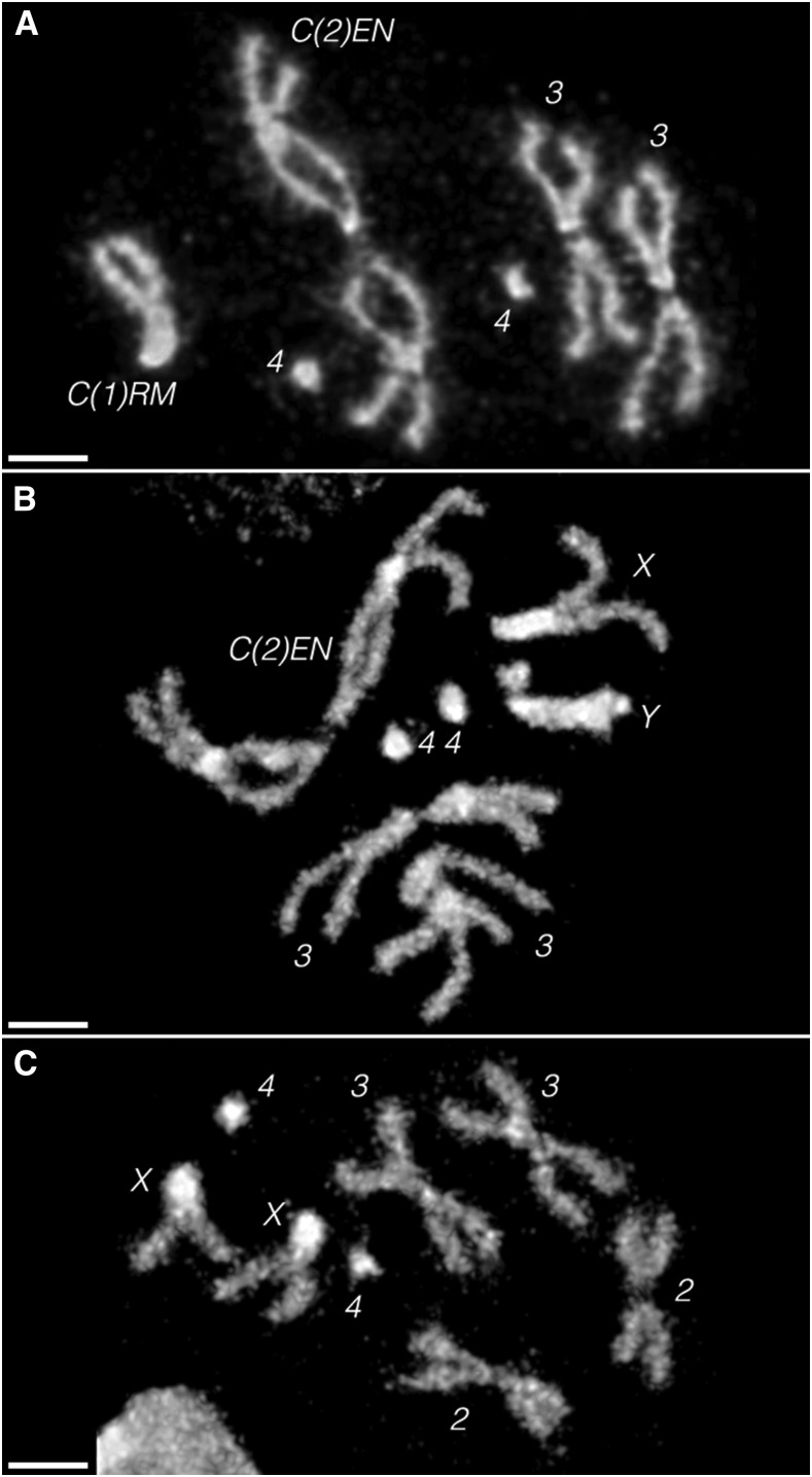
Figure 2.
Brain squashes of (A) a C(1)RM, y2 su(wa) wa/Ø; C(2)EN, bw sp/Ø female third instar larva, showing only six chromosomes, (B) a X/Y; C(2)EN, bw sp/Ø male larva, showing seven chromosomes and (C) an Oregon-R female larva, showing eight chromosomes. The constrictions in the middle of the arms of C(2)EN match previously published images of this chromosome in mitosis (Martins et al. 2013). These DAPI (4’,6-diamidino-2-phenylindole) images are representative of the karyosomes seen in at least 10 larvae of each sex. Note the lack of any free Y in the female, and that the sex chromosomes in the male are clearly not attached, indicating that males must undergo X/Y ⇔ C(2)EN segregation. All scale bars, 2 µm.

