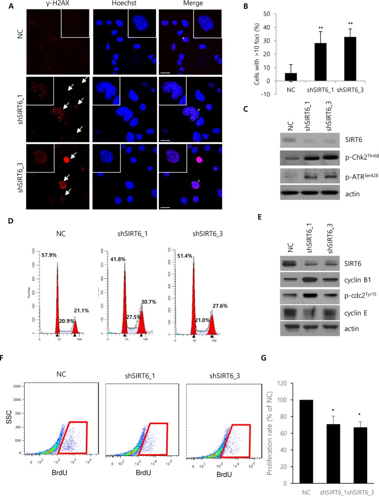
Fig 5. Effect of SIRT6 knockdown on DNA damage and cell cycle progression.
(A) Expression of γ-H2AX was assessed by immunocytochemistry using a confocal microscope. (B) Cells with over 10 foci were quantified by counting cells from the randomly obtained images. The quantified cells were compared in the histogram. The scale bar indicates 30 μM. (C) The expression of indicated proteins was assessed by western blotting. Cells for western blotting and immunocytochemistry were harvested or fixed 5 days after viral transduction. (D) Propidium iodide staining was performed to determine the cell cycle distribution in Hep3B cells 5 days after infection with lentiviruses containing NC or shSIRT6. The numbers of Hep3B cells at the G1, S, and G2/M phases were quantified by FACS analysis. The Modifit program was used for data analysis. (E) Expression of cell cycle-related proteins and SIRT6 were assessed by western blotting. Cells for microarray, RT-PCR, and western blotting were harvested 5 days after viral transduction. All data are representative of three independent experiments. (F) Representative FACS analysis of BrdU incorporation. (G) Cell proliferation rate of NC and shSIRT6-depleted cells as quantified by FACS analysis of four independently performed experiments. (**P<0.01, *P<0.05).

