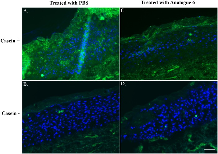
Fig 7. In-Situ Zymography in Organotypic Skin Culture (OTC).
Fluorescence images from in-situ zymography of OTC treated with PBS (A & B) or Analogue 6 (C & D) are shown. For the negative staining, PBS was applied to the cryosection instead of casein substrate solution (B & D). Cell nuclei were counterstained with 4',6-diamidino-2-phenylindole (blue color) and protease activity is signaled by green fluorescence. All images were obtained in MicroManager (Ver.1.4.21) with an exposure time of 1s and images were processed using ImageJ (Ver.1.46) and finalized by Adobe Photoshop CS. Brightness and contrast settings were all the same and applied to the entire images. Scale Bar = 200 μm.

