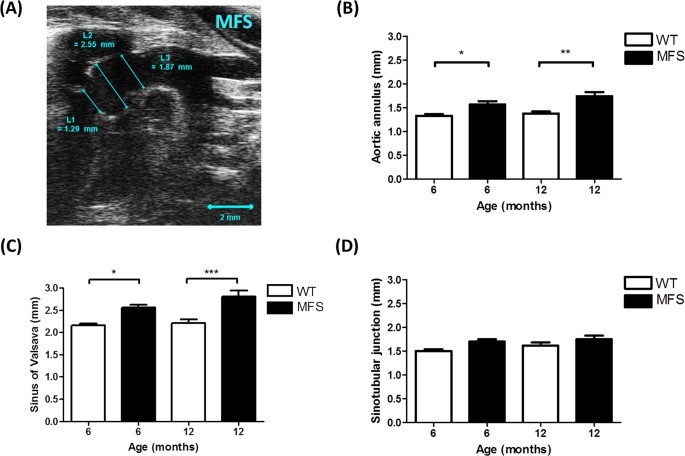
Fig 1. Aortic root dimension of WT and MFS mice.
(A) B-mode view of the aortic arch from a 6-month MFS mouse. Diameters of the (B) aortic annulus [L1], (C) sinus of Valsalva [L2] and (D) sinotubular junction [L3] were significantly increased in MFS mice versus WT. The larger aortic root diameter indicates significant aortic dilation. * indicates p < 0.05, ** indicates p< 0.01, *** indicates p< 0.001. (n = 8)

