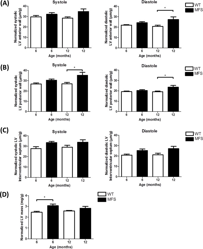
Fig 4. Echocardiographic assessment of left ventricle (LV) mass and wall thickness.
Left ventricle (LV) mass, systolic and diastolic wall thickness were normalized by body weight (g). Data presented are normalized systolic and diastolic (A) anterior wall thickness, (B) posterior wall thickness, (C) interventricular septal thickness and (D) normalized LV mass from WT and MFS mice (n = 8). * indicates p< 0.05.

