
Fig. 1.
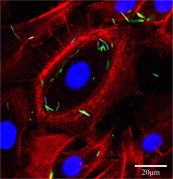
Intracellular localization of Fusobacterium nucleatum in GECs. Immunofluorescence confocal micrograph GECs infected with F. nucleatum (MOI of 100) for 1 h. The single optical section through the middle of the host cell confirms the intracellular localization of the bacteria. Fixed GECs were stained with phalloidin–tetramethylrhodamine B isothiocyanate (red) to show actin filaments, and anti-F. nucleatum antibody (green). Bar represents 20 μm.