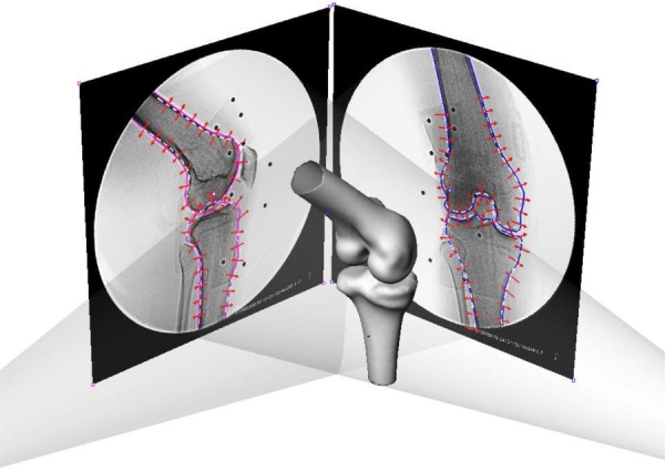
Fig. 2.

Environment setup for the SSM in a virtual dual fluoroscope imaging system. Avg models, including the femur and tibia, were imported and positioned to match the silhouettes on the dual fluoroscopic images. Corresponding SSM would deform to fit the outlines of dual-fluoroscopic images.
