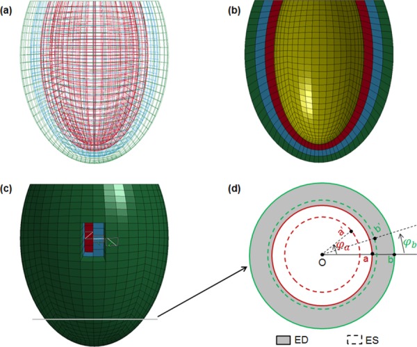
Fig. 1.

A 3D FE model was created to simulate the function of LV. (a) Wireframe view of the LV, which was represented by an ellipsoidal morphology; (b) view with half of the model removed to show the LV wall with three transmural layers of equal thickness, outermost = epicardium, middle = midwall, and innermost = endocardium; (c) epicardial view of model with transmural variation in myofiber orientation (white lines within elements) and a radial–circumferential plane near the apex where the LV twist was assessed, and (d) example of the rotation angles of two specific nodes on the inner and outer surface of LV wall from end-diastolic to ES states.
