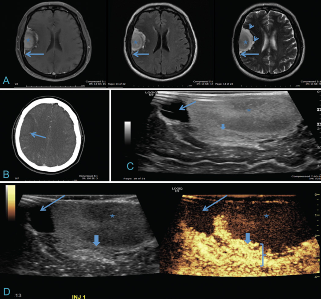
Fig. 3.
Meningioma with corresponding iCEUS images. A: Axial T1-weighted contrast-enhanced, FLAIR, and T2-weighted MR images of a right frontotemporal, dura-based, extraaxial lesion. The core of the lesion is mildly heterogeneous (asterisk) with a dural tail (long arrows) and cystic components seen on T2-weighted images (short arrows). B: Axial CT angiogram demonstrating a feeding vessel from the middle cerebral artery distribution (arrow). C: Intraoperative ultrasound image demonstrates a cystic component (long arrow) and heterogeneous echotexture within the core of the lesion (asterisk) and a thick heterogeneous peripheral component (thick arrow). D: Corresponding B-mode (left) and iCEUS (right) images demonstrating a central tumor with low vascularity (asterisk), nonenhancing cystic component (long arrow), and thick, avidly enhancing peripheral rim (thick arrow and bracket). Differentiation can be appreciated between enhancing tumor and normal, adjacent brain parenchyma.

