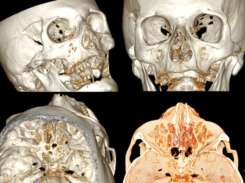
Fig. 2.

3D CT scan reconstruction showing four different projections of frontosphenotemporal Pellerin et al fracture pattern; note in frontal view (up right), SOF size reduction caused by medial displacement of the entire right lateral orbital wall; the black dashed line in the intracranial view (down right) shows the medial collapse of lateral orbital wall into the SOF. CT, computed tomography; SOF, superior orbital fissure; 3D, three-dimensional.
