
Figure 2. Strategies that induce HIF-2α are sufficient to suppress efferocytosis.
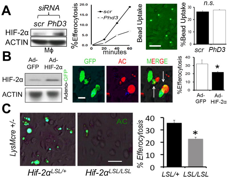
(A) Bone marrow-derived Mϕs were treated with scrambled (scr) vs. Phd3 siRNA and HIF-2α levels revealed by immunoblot, as a function of efferocytosis and engulfment of control latex beads. (B) Adeno-HIF-2α-GFP (green) was transduced into Mϕs and immunoblots and efferocytosis of red apoptotic cells (ACs) quantified. (C) Non-stained primary Hif-2α LSL/+ LysMcre vs. Hif-2α LSL/LSL LysMcre Mϕs were challenged with ACs and efferocytosis imaged and quantified. Scale bar = 60 micrometers. * indicates < 0.05 relative to control.