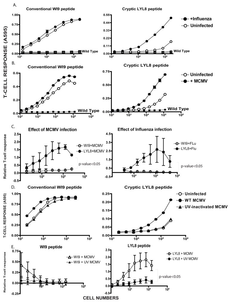
Figure 4. Cryptic peptide presentation is enhanced by virus infection.
A. Primary macrophages from the WI9.LYL8 transgenic mice were infected with Influenza virus at an MOI of 1.0 for 6 hours, after which the cells were harvested and assayed for antigen presentation activity using T-cell hybridomas as described in Figure 2D. The Y-axis indicates the T-cell response (11p9Z hybridoma response for the conventional WI9 peptide and the BCZ103 hybridoma response for the cryptic LYL8 peptide) and X-axis shows the the cell numbers per well. Data is representative of 3 experiments. B. WI9.LYL8 primary macrophages were infected with Mouse-cytomegalovirus (MCMV) at an MOI of 1.0 for 6 hours. Cells were then harvested and a T-cell hybridoma assay was set up as described in Figure 2D. The Y-axis indicates the T-cell response (11p9Z specific hybridoma response for the conventional peptide and the BCZ103 specific hybridoma response for the cryptic peptide) and X-axis indicates the cell numbers per well. Data is representative of 4 experimental repeats C. T-cell responses to the WI9 and LYL8 peptides, upon MCMV infection (left panel), Influenza infection (right panel), were normalized to that of the uninfected samples for 3 distinct experiments. An unpaired t-test (with Welch’s correction) was performed, comparing the conventional and cryptic peptide responses - p-value<0.05 D. WI9.LYL8 macrophages were infected with wild-type MCMV or UV-inactivated MCMV for 6 hours. Cells were then harvested and a T-cell hybridoma assay was set up as described in Figure 2. The Y-axis indicates the T-cell response (11p9Z specific hybridoma response for the conventional peptide and the BCZ103 specific hybridoma response for the cryptic peptide) and X-axis indicates the cell numbers plated. Data is representative of 3 experiments. E. T-cell responses to the WI9 (left panel) and LYL8 (right panel) peptides upon wild-type and UV-inactivated MCMV infection, were normalized to that of the uninfected samples for 3 distinct experiments. An unpaired t-test (with Welch’s correction) was performed, comparing the wild-type and UV-inactivated T-cell responses.

