
Fig. 2.
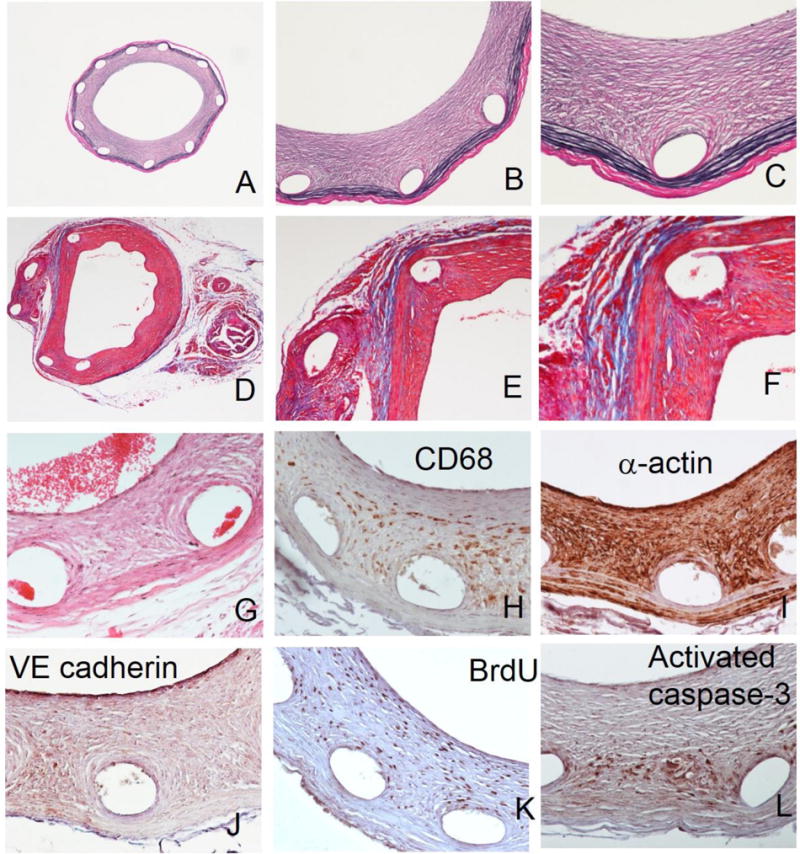
Special staining and immunohistochemistry of paraffin sections prepared following metallic stent dissolution.
Representative arterial sections stained according to Verhoeff-van Gieson (A–C), Masson’s trichrome (D–F) and H&E (G) protocols. Typical results of immunohistochemical staining with primary antibodies chosen to detect CD68-positive macrophages (H), α-actin-positive SMC (I), VE cadherin-positive endothelial cells (J), proliferating BrdU-positive cells (K) and activated caspase 3-positive cells undergoing apoptosis (L). Original magnifications: 2A, 2D – 40x; 2B, 2E – 100x; 2C, 2F–2L – 200x.