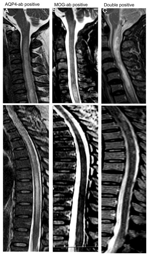
Figure 5.
Spinal lesions in NMOSD patients with different antibody profiles during attacks. A, Sagittal T2 weighted images demonstrating single typical longitudinally extensive T2 hyperintense, central and severer edema spinal cord lesion from the cervical to thoracic cord of a patient with AQP4-ab. B, Sagittal T2 weighted images of multiple spinal lesions in a patient with MOG-ab: longitudinally extensive T2 hyperintensity and mildly edematous spinal cord lesion affect the cervical cord. Multiple discontinuous, short-segments, patchy signal abnormalities affect the thoracic cord to conus area. C, Sagittal T2 weighted images of multiple spinal lesions in a double-positive patient: longitudinally extensive T2 hyperintense, central and severe edematous spinal cord lesion affects the cervical cord and multiple discontinuous, short-segment, patchy signal abnormalities affect the thoracic cord to conus area.

