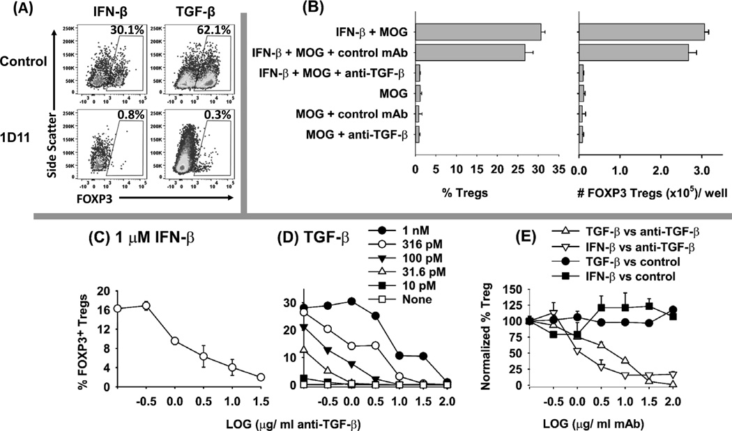
Figure 10. The anti-TGF-β mAb 1D11 inhibits IFN-β dependent induction of FOXP3+ Tregs in vitro.
(A–E) Naïve 2D2-FIG splenocytes (200,000/well) were cultured in triplicate with 1 µM MOG35-55 and either 1 µM IFN-β or 100 pM TGF-β (or as designated in D). Cultures also included either anti-mouse-TGF-β (1D11) or the isotype control mAb (LRTC 1) (31.6 µg/ ml) (A–B) or as designated on the x-axis (C–E). After 7 days of culture, CD4+ T cells were assessed for GFP expression. (A) Shown are representative dot plots of side scatter (y-axis) and FOXP3 expression (x-axis) together with (B) percentages and absolute numbers of FOXP3+ Tregs after culture with MOG35-55 in the presence or absence of IFN-β and mAb. The quantitative neutralization profiles are shown for the anti-TGF-β 1D11 mAb in cultures of IFN-β-induced Tregs (C, E) and TGF-β-induced Tregs (D, E). Error bars represent standard error of the mean. These data are representative of three independent experiments.

