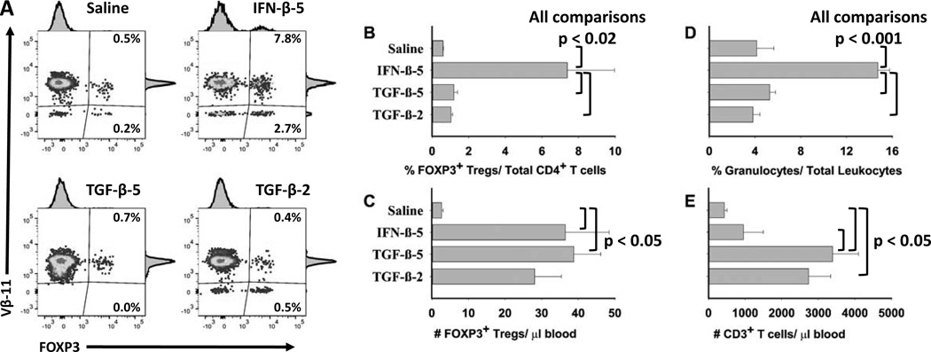
Figure 11. When combined with Alum and MOG35-55, IFN-β uniquely increased the percentages of FOXP3+ Tregs relative to the total pool of CD4+ T cells.
On day 0, 2D2-FIG mice (n = 5) were injected with “Saline in Alum”, “5 nmoles IFN-β + 5 nmoles MOG35-55 in Alum” (IFN-β-5), “5 nmoles TGF-β + 5 nmoles MOG35-55 in Alum” (TGF-β-5), or “2 nmoles TGF-β + 5 nmoles MOG35-55 in Alum” (TGF-β-2). On day 13, PBMC were assessed for FOXP3+ T cells as a percentage of the CD45+, CD3+ CD4+ T cell population (A–B), the total numbers of FOXP3+ Tregs per µl of blood (C), the percentages of granulocytes as a percentage of total leukocytes (D), and the total numbers of CD3+ T cells per µl of blood (E). Analysis panels included CD3-BV421, CD4-PE, Vβ11-AF647, and CD45-BV785.

