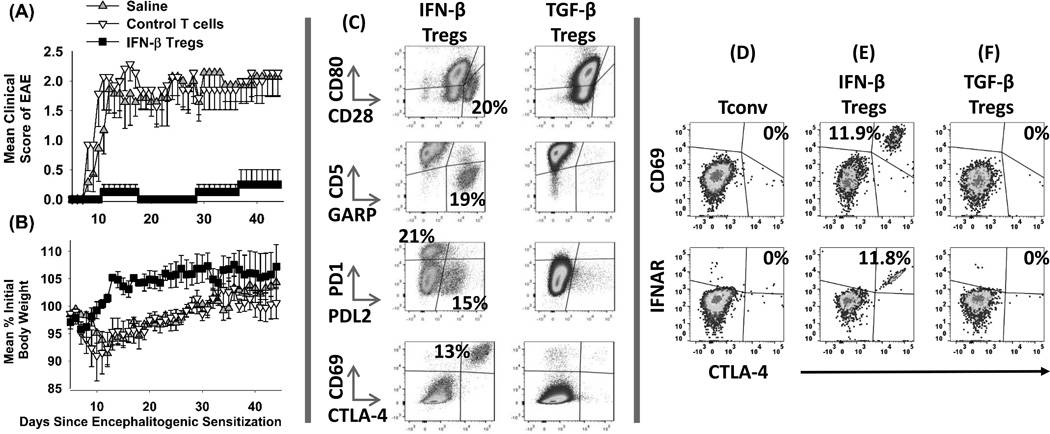
Figure 5. Function and phenotype of IFN-β-induced Tregs.
2D2-FIG splenocytes were cultured with 1 µM MOG35-55 and IL-2 in the presence (IFNβ-Tregs) or absence (Control T cells) of 1 µM IFN-β for 7 days. Donor IFNβ-Tregs and control T cells were extensively washed after the 7-day culture and injected at a dose of 107 total T cells on day 4 into recipients that were previously challenged with MOG35-55/ CFA (day 0) and Pertussis toxin (days 0, 2). Error bars represent standard error of the mean. Shown are 1 of 2 experiments that were compiled in Table 1. (C–F) 2D2-FIG T cells were activated with 1 µM IFN-β + MOG35-55 (C & E, FOXP3+ Treg gate), 1 nM TGF-β + 1 µM MOG35-55 (C & F, FOXP3+ Treg gate), or 1 µM MOG35-55 alone (D, Tconv cell gate) for 7 days. T cells were analyzed for the designated surface markers. These data are representative of three independent experiments. Cells were stained with CD45-BV785 and CD3-PE.CF594 within different panels that included; [CD86-BV421, CD80-PE, and CD28-APC], [CD69-BV421, CD5-PE, GARP-AF647, and PDL1-PE.Cy7], [PD1-BV421, PDL2-PE, and PDL1-APC], and [CD69-BV421, CTLA4-PE, and IFNAR1-APC].

