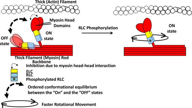
Fig. 3.
Graphical representation of the effect of RLC phosphorylation on myosin head and lever arm arrangement. (Figure modified from Fig. 6c of Farman et al. 2009) Model based on skeletal muscle (Duggal et al. 2014; Midde et al. 2013) and cardiac muscle (Kampourakis and Irving 2015). In this model, without RLC phosphorylation, the myosin S1 and RLC regions are in conformational equilibrium between lying/binding to the thick filament backbone surface (OFF state) and moving away towards thin filament (ON state) controlled by the RLC and the thick filament surface. This equilibrium is shifted towards the ON state upon RLC phosphorylation, possibly due to weakened electrostatic interaction between the added phosphate and negatively charged thick filament surface. The entire thick filament arrangement of myosin heads and other sarcomeric proteins (MyBP-C, Troponin complex etc.) are not shown for simplicity. Not drawn to scale. (Notes: The ON and OFF states mentioned here are not related to the activation/deactivation with calcium binding/unbinding to Troponin C. The OFF-state myosin heads without RLC phosphorylation is drawn here lying on the thick filament backbone in the same direction as in the ON state, but it is possible that the OFF-state heads may fold on the opposite direction interacting with the thick filament backbone as drawn previously in Fig. 5 of Kampourakis and Irving 2015)

