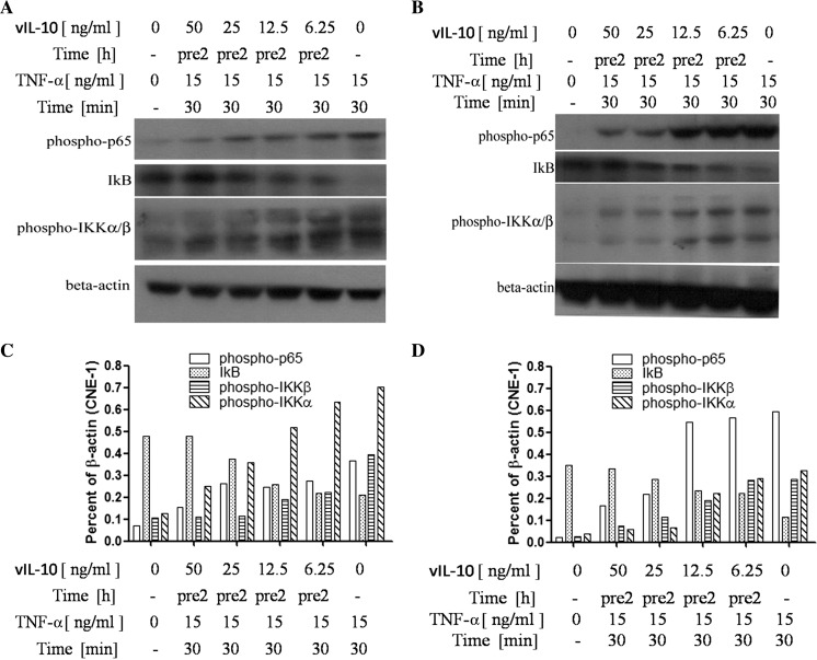
Fig. 1.
The activity changes of the NF-κB signaling pathway in NPC cells after treatment with vIL-10. a The CNE-1 cells were treated with TNF-α (15 ng/ml) for 30 min or untreated (−). Alternatively, the cells were treated with vIL-10 for 2 h (pre2) before TNF-α treatment. The cells were lysed for Western blot analysis as described in the “Materials and methods” section. b CNE-2 cells were treated in the same manner as a, and the results are presented. c The results in a were semi-quantified by ImageJ. d The results in b were semi-quantified by ImageJ

