
FIG 2 .
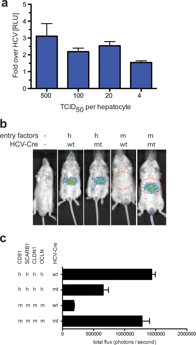
mtHCV can enter mouse hepatocytes in vitro and in vivo. R26-LSL-Fluc mice were injected with 1011 adenoviral particles encoding human (h) or mouse (m) CD81, SCARB1, CLDN, and OCLN. Bioluminescence was measured at 72 h postinjection of 2 × 10⁷ 50% tissue culture infectious doses (TCID50) of cell culture-produced murine tropic (mt) or wild-type (wt) HCV-Cre. (a) Primary hepatocytes isolated from C57BL/6 mice were plated on collagen-coated plates, and monolayers were infected with Jc1(p7nsGluc2A) or Jc1/mCD81Gluc2A with the indicated numbers of infectious particles (as titered on Huh-7.5 cells) per primary hepatocyte. Luciferase activity was measured 48 h postinfection. Data are represented as the signal of mtHCV over the parental strain. (b) Representative pseudo-colored images. (c) Quantitation of total photon flux/second (n ≥ 4). Values are means plus standard deviations (SD) (error bars).