Figure 5.
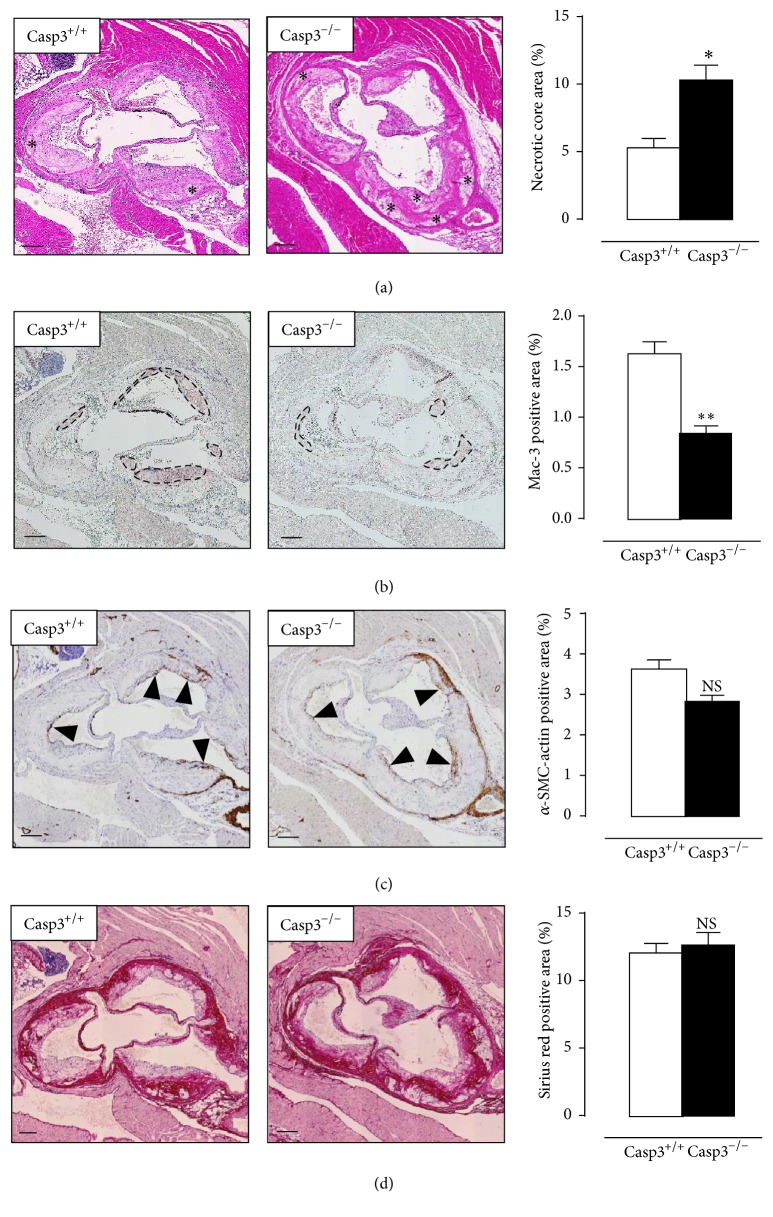
Caspase-3 deficiency increases plaque necrosis in ApoE−/− mice. Casp3+/+ApoE−/− mice (Casp3+/+) (n = 8) and Casp3−/−ApoE−/− mice (Casp3−/−) (n = 7) were fed a western-type diet for 16 weeks. (a) Aortic root atherosclerotic plaques were stained with H&E to quantify plaque necrosis (the necrotic cores are indicated by an asterisk). (b, c) Serial sections were immunostained for Mac-3 and α-SMC-actin to determine macrophage content (Mac-3 positive regions are indicated by black circles) and vascular smooth muscle cell content (α-SMC-actin positive fibrous caps are indicated by black arrowheads), respectively. (d) Serial sections were stained with Sirius red to quantify total collagen content (characterized by intense pink/red staining). (∗ P < 0.05, ∗∗ P < 0.01, and NS, not significant, versus Casp3+/+; repeated measures) scale bar: 150 μm.

