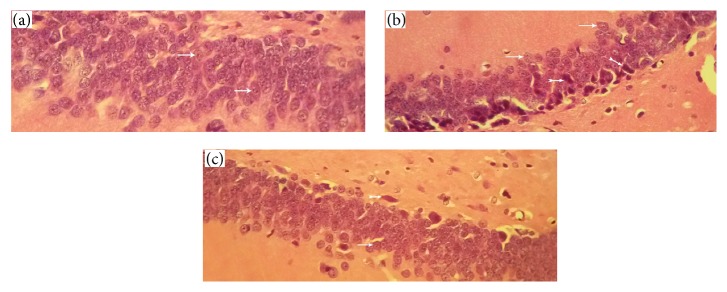
Figure 6.
Histopathological assessment. (a) Control group neurons have an integrity and regular structure with normal morphology, round nuclei, prominent nucleolus, and clear cytoplasm. (b) Ischemia group neurons have numerous pyramidal cells with pyknotic nuclei and lack of nucleolus and hyperchromatic cytoplasm. (c) Treatment group have considerably decreased numbers of cells with pyknotic nuclei and lack of nucleolus and hyperchromatic cytoplasm compared to the ischemia group (thin arrows indicate normal cells and thick arrows indicate apoptotic cells).

