Figure 1.
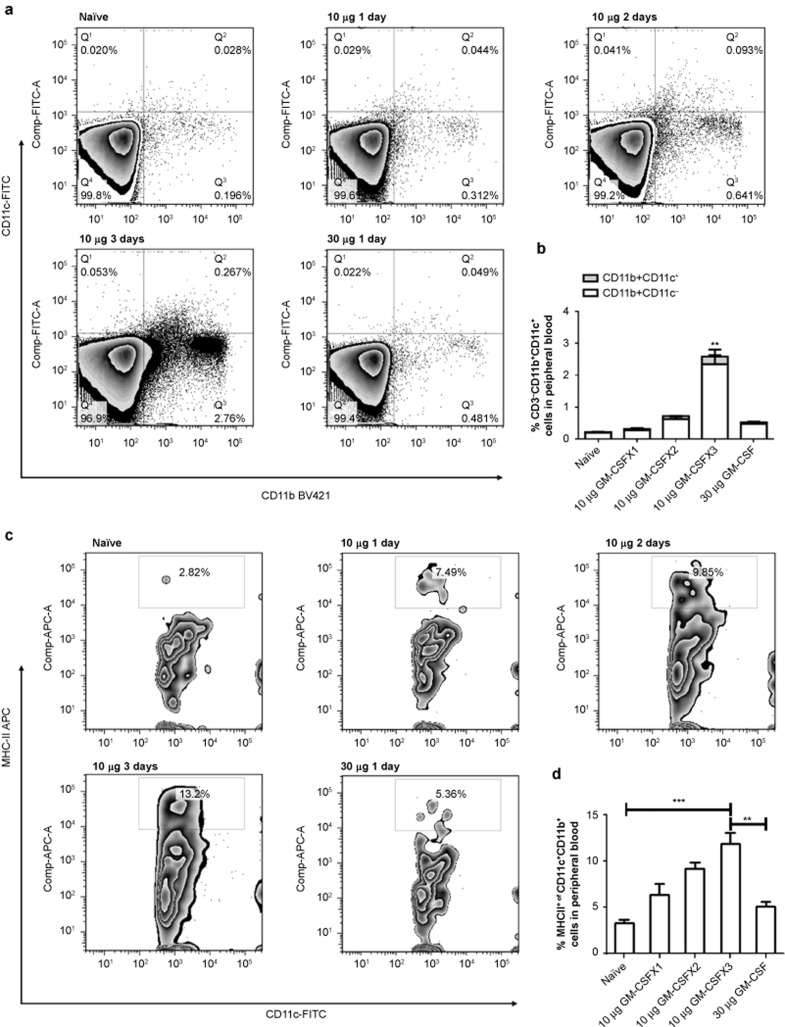
GM-CSF stimulation exerts time-sensitive effects on dendritic cells in vivo. HBV transgenic mice were subcutaneously injected with 10 μg of GM-CSF daily for one, two, and three days or with 30 μg of GM-CSF for one day. Peripheral blood cells were isolated on day 4. FACS analysis was performed to determine the percentage of DCs with a myeloid phenotype by staining with CD11c-FITC, CD11b-PB, and MHC-II-PE. The percentages of double-stained cells were calculated. The CD11c+CD11b+ cells among peripheral blood cells (Figure 1a and 1b) were immunostained for MHC-II (Figure 1c and 1d) and analyzed by FACS. Data are shown as mean ± SEM (n = 3) and represent one of the three independent experiments. *p <0.05, **p <0.01, and ***p <0.005 (unpaired Student's t-test).

