Figure 4.
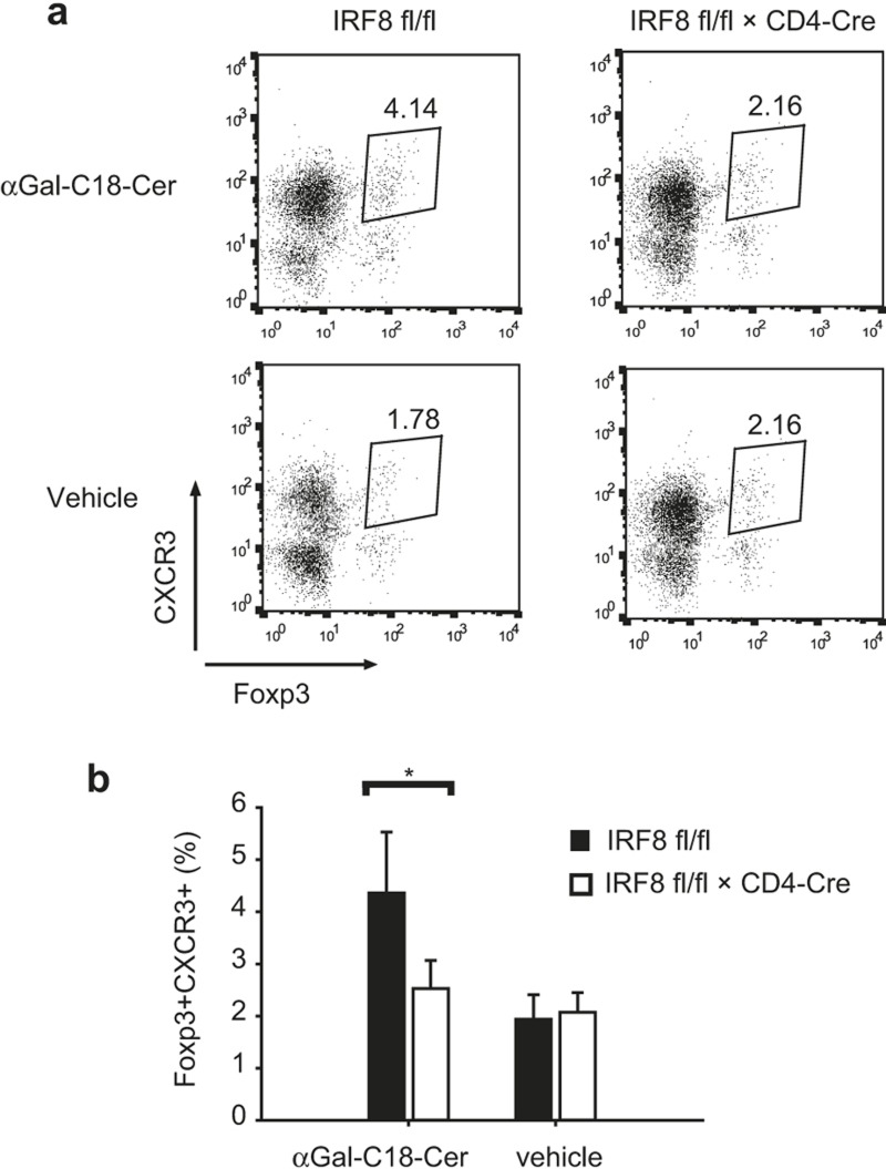
Defective recruitment of CXCR3+ Treg cells to an inflammation site. (a) Wild-type or CD4-specific IRF8-deficient mice were injected with αGal-C18-Cer or vehicle control intraperitoneally. After one day, CD4 T cells were isolated from the liver and the expression of Foxp3 and CXCR3 was analyzed by FACS. Data are representative of three independent experiments with similar results. (b) Ratio of Foxp3+CXCR3+ cells in CD4 T cells from the liver. Three experiments of (a) were combined. The error bar shows standard deviation (n = 3∼8). Statistical difference was analyzed by Student's t-test. *p < 0.05.

