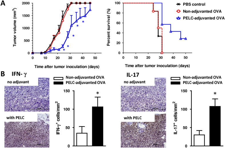
Figure 5. Immunotherapeutic effect of the OVA protein adjuvanted with PELC and administered to C57BL/6 mice bearing EG7 tumor cells.
(A) Anti-tumor efficacy; (B) IHC analysis of IFN-γ and IL-17 expression at tumor-adjacent counterparts. Mice were inoculated s.c. in the flank with EG7 tumor cells (2 × 10⁵ cells/mouse). Upon the appearance of palpable tumors, six mice per group were injected s.c. at the tail base with 10 μg/dose OVA protein with or without PELC on day 7. Mice were euthanized when the tumor volume exceeded 2,000 mm3. The data are expressed as the mean value ± standard deviation. The tumor volumes were compared on days 21, 24, 28, 31 and 35 following the onset of tumor growth in the vaccine group. On day 31 after tumor cell inoculation, tissue sections from tumor-adjacent counterparts (n = 3) were IHC stained with IFN-γ and IL-17 antibodies (original magnification, ×400). Cells with brown signals around the blue nuclei indicate IFN-γ+ or IL-17+ cells. Statistical significance was determined by performing the two-tailed Student’s t-test. *P < 0.05 compared with the non-adjuvanted OVA group. The data are representative of two independent experiments.

