
Figure 6. The role of mononuclear phagocytes during endometrial repair.
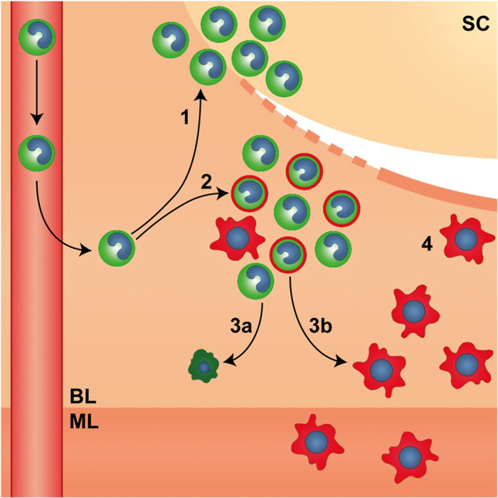
Endometrial repair is rapid and occurs concurrently with tissue breakdown and clearance of shed cells (SC). Endometrial repair is associated with a dynamic influx of ‘classical’ monocytes from the circulation in response to the ‘wounding’ stimulus of menses which differentiate in response to spatially distinct signals within the tissue. 1. GFP+ monocytes (green) cluster in areas of tissue breakdown and shedding. 2. A mixed population of GFP+ ‘classical’ monocytes and GFP+F4/80+ monocyte-derived macrophages (green/red) cluster in areas of active repair and remodelling in close proximity to denuded stromal surfaces (dotted line). 3a. GFP+ monocytes may undergo apoptosis and be cleared from the tissue following resolution of inflammation or 3b. undergo differentiation into tissue resident macrophages. 4. F4/80+ tissue resident macrophages (red) are associated with newly re-epithelialized areas of repaired tissue (solid line) and are detected within the basal (BL) and myometrial layer (ML) of the uterus.