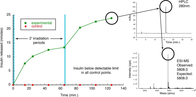
Figure 3.
In vitro insulin PAD photolysis. PAD material was exposed to two 2′ periods of 365 nm LED light (blue bars). Supernatant was monitored for insulin release (left). Material released showed a retention time in HPLC consistent with insulin (upper right), and this was confirmed to be insulin via ESI-MS (lower right).

