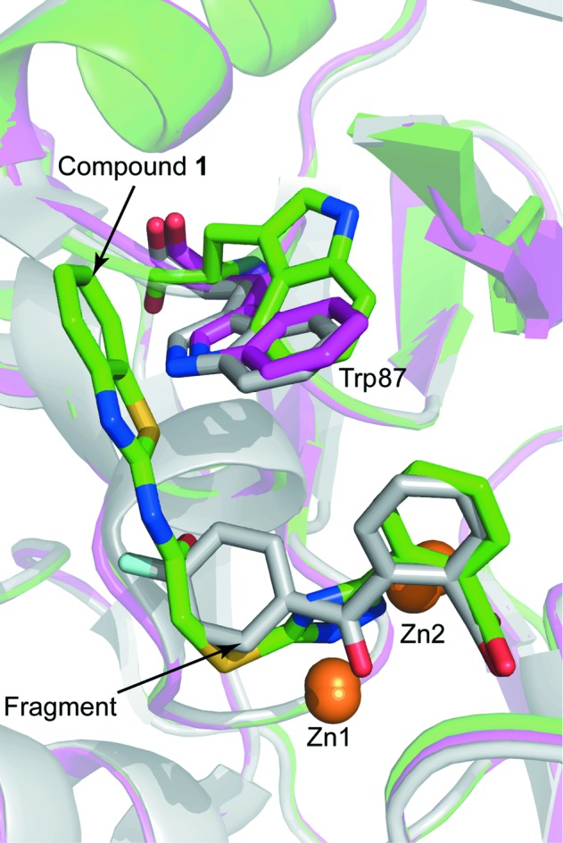
Figure 6.

Structural alignment of a native VIM-2 structure (purple; PDB entry 5acu; Christopeit et al., 2015 ▸), the VIM-2 structure in complex with compound 1 (green) and the VIM-2 structure in complex with a fragment (grey; PDB entry 5acx; Christopeit et al., 2015 ▸). The protein backbones are shown as ribbon cartoons. Zinc ions are shown as orange spheres and Trp87, compound 1 and the fragment are shown as sticks.
