Figure 1.

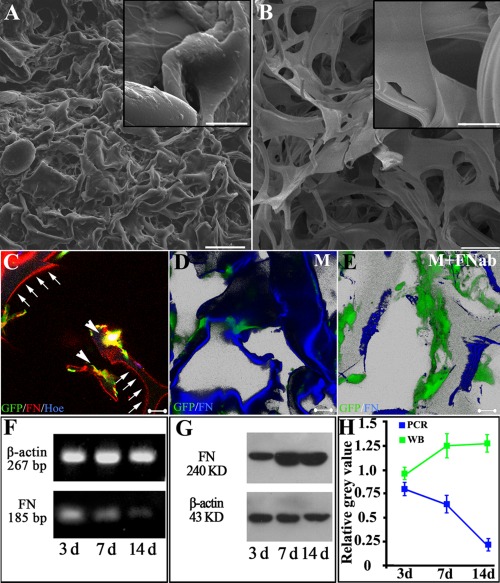
Fibronectin deposition on the surface of gelatin material after co‐culture of MSCs and SCs. ECM accumulation is significant in gelatin sponge (GS) in the MSCs group (A), as evidenced by the rough surface (inset in A), while the surface of GS without MSCs is smooth (inset in B). FN, secreted by GFP positive MSCs (arrowheads in C), deposits onto the surface of gelatin material and displays thread‐like red fluorescence (arrows in C). Application of FN antibody decreases the deposition of FN onto the surface of gelatin material. Three dimensional reconstructive image shows that the surface of gelatin material is decorated by adherent FN (blue, in D). After adding FN blocking antibody (FNab) to the culture medium, deposition of FN (blue) onto the surface is obviously reduced as shown in (E). Green cells in D and E are MSCs. RT‐PCR and Western blot results indicate that, although the transcriptional level of FN decreases with the increase in duration of co‐culture (F), the amount of FN protein increases within the gelatin sponge (GS) scaffolds (G,H). Scale bars: 200 μm in (A,B), 20 μm in the inset of (A,B), and 20 μm in (C–E).
