
Figure 4.
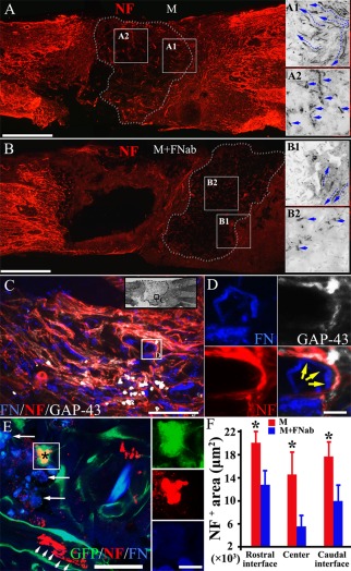
Autocrine FN increases nerve fiber regeneration after spinal cord injury. Scaffolds containing MSCs with or without FN blocking antibody treatment for 14 days of culture are transplanted into the injured spinal cord for 4 weeks. NF positive nerve fibers (red), which are bulky and of varying lengths, are observed in the M group (A). These regenerating nerve fibers grow preferentially toward the implant (A1). NF positive fibers are common and are readily identified in the center of the implant (A2). When implanted with FN blocking antibodies treated scaffold, NF positive nerve fibers (B) are uncommon and often in disarrays (B1). They are rarely seen in the center (B2) of the implant in the M + FNab group when compared with the same area (A2) of the M group. Autocrine FN was detected at 4 weeks after implantation, as indicated by the presence of many thread‐like fluorescent short fibers in the injury/graft site of spinal cord (C). Nerve fibers attached to FN fibers are common (yellow, arrows in D), with some of them being GAP‐43 positive, suggesting robust nerve fiber regeneration in the FN enriched area (C,D). MSC‐derived neuron‐like cells (red, asterisk of E) can be found inside the implant, surrounded by a significant group of GFP positive donor cells expressing FN (blue). There are a few NF positive nerve fibers (red, arrowheads in E) near the donor cells (GFP positive, in E). FN accumulation in the extracellular space is observed inside the implant (arrows in E). Histogram chart shows a prominent increase of innervation at the rostral interface between the host tissue and implant, the center of the implant and the caudal interface between the host tissue and implant in the M group, compared with the M + FNab group. Asterisks in (F) indicate p < 0.05 (n = 8, Student t‐test). Scale bars: 1 mm in (A) and (B); 200 μm in (C); 20 μm in (D); 50 μm in (E); 10 μm in inset of (E).