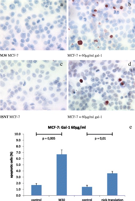
Fig. 3.

M30 staining in untreated for MCF-7 cells had a mean of 1.7 % (a). In cells treated with 60 μg/ml gal-1 for 48 h the rate of very early apoptosis is elevated to up to 6.7 % (p = 0.005, b). The normal rate of apoptosis in MCF-7 breast cancer cells had a mean of 1.4 % detected by in situ nick translation (ISNT, c). The incubation with 60 μg/ml gal-1 for 48 h significantly enhanced apoptosis in MCF7-cells to a maximum of 3.6 % (p = 0.01, d). Results of M30 and ISNT staining are summarized (e) (n = 10)
