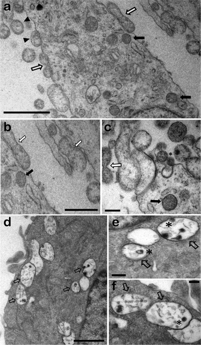
Figure 2. Transmission electron microphotographs of olfactory ensheathing cells (OECs) (a–c) and microglia (d–f) infected with Streptococcus pneumoniae.

(a) An overview of different invasion stages, showing adherent pneumococcal cells associated with electron-dense material (arrowheads) and diplococcus-shaped pairs (white arrows), as well as intracellular pneumococci in vacuoles (black arrows). (b) Subsequent engulfment by cellular protrusions, with the formation of a membrane-bound vacuole. (c) Asterisk indicates a possible clathrin-coated pit-like structures and electron-dense material associated with invading bacteria. (d–f) Intracellular pneumococci in vacuoles showing clear signs of degradation. Higher-magnification views of digested bacteria in cytosolic vacuoles (asterisk in e,f). Scale bar = 1 μm (a); 0.7 μm (b); 0.2 μm (c); 1 μm (d); 0.2 μm (e); 0.2 μm (f).
