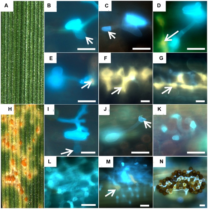
FIGURE 1.

Development of fungal structures in the resistant accession PI272560 (A–G) and the susceptible accession 36554 (H–N). Macroscopic symptoms on leaf segments 168 hai (A,H) and microsopic observations 6 hai (B,I), 12 hai (C,J). 24 hai (D,K), 48 hai (E,L), 72 hai (F,M), and 168 hai (G,N) are compared. Arrows in (B,I) mark the substomatal vesicle, in (C) the haustorial mother cell and in (D,E,F,G,M) autofluorescence around infection sites. Bars: 20 μm.
