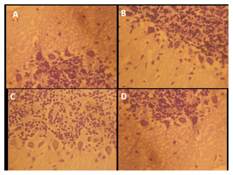
Figure 10.
Representative photo micrograph of the sections of the cerebellar cortices following saline, cannabis, NSO and cannabis+NSO administrations showing: normal nissl distributions in the soma and unaffected proximal dendrites of the large cerebellar Pukinje neurons in A and C; contrasting stains, pyknotic and darkly stained cell body, presence of dark neurons and vacuolation in the surrounding neuropil of Pukinje cells in B; marked regeneration, less shrunken cells. (CFV × 400)

