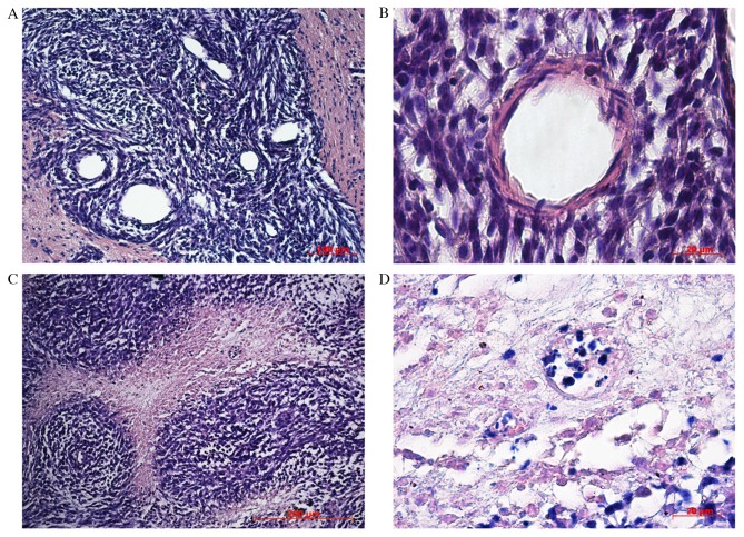
Figure 4.
Hematoxylin and eosin staining of rat brain tumors, 20 days after implantation of tumor cells. (А) Tumor tissue with newly formed blood vessels, (B) Blood vessel development among satellite tumor cells. (C) Angiocentric clusters of tumor cells. (D) Necrotic areas in tumor tissue, 30 days after implantation of tumor cells.

