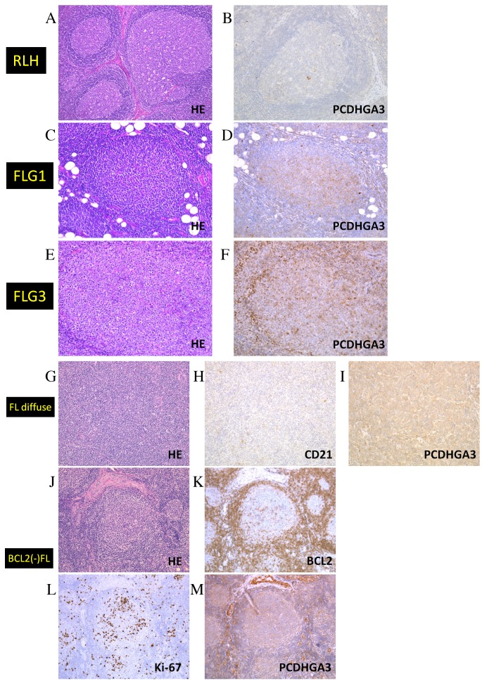
Figure 2.
Immunohistochemical analysis of PCDHGA3 in FL samples. (A) HE staining of RLH. (B) PCDHGA3 was negative in RLH. (C) HE staining of an FL grade 1 sample, which was (D) positive for PCDHGA3. (E) HE staining of an FL grade 3 sample, which was (F) positive for PCDHGA3. (G) HE staining of an FL with diffuse area sample, which was (H) negative for CD21-expressing follicular dendritic cells and (I) positive for PCDHGA3. (J) HE staining of a BCL2-negative FL sample, which was (K) negative for BCL2, (L) positive for Ki-67 and (M) positive for PCDHGA3. All these images were representative of their groups. Original magnification A and G-M, ×100. Original magnification B-F, ×200. PCDHG, protocadherin γ; FL, follicular lymphoma; HE, hematoxylin and eosin; RLH, reactive lymphoid hyperplasia; CD, cluster of differentiation; BCL2, B-cell lymphoma 2.

