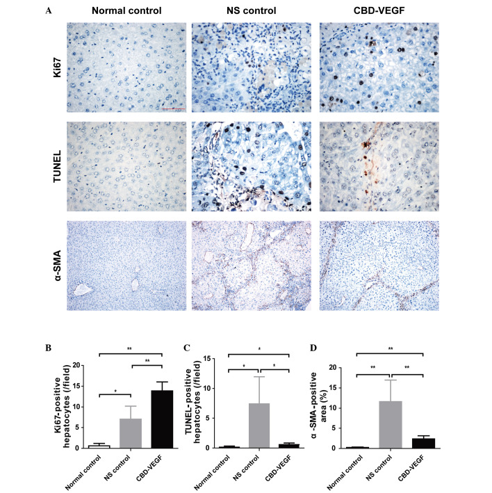
Figure 4.
CBD-VEGF promoted hepatocyte proliferation and suppressed hepatocyte apoptosis and HSC activation. Immunohistochemical staining of Ki67 and α-SMA in addition to a TUNEL assay were performed in the liver sections of each group. (A) Upper panels, Ki67 immunohistochemical staining (magnification, ×400); middle panels, TUNEL assay for evaluation of hepatocyte apoptosis (magnification, ×400); lower panels, α-SMA immunohistochemical staining (magnification, ×100). (B) Statistical analysis of proliferative hepatocytes, (C) TUNEL-positive hepatocytes and (D) α-SMA positive area of each group. n=5 for each group. Data are presented as the mean ± standard deviation; **P<0.01, *P<0.05. CBD-VEGF, collagen-binding domain-vascular endothelial growth factor; HSC, hepatic stellate cells; α-SMA, α-smooth muscle actin; NS, normal saline.

