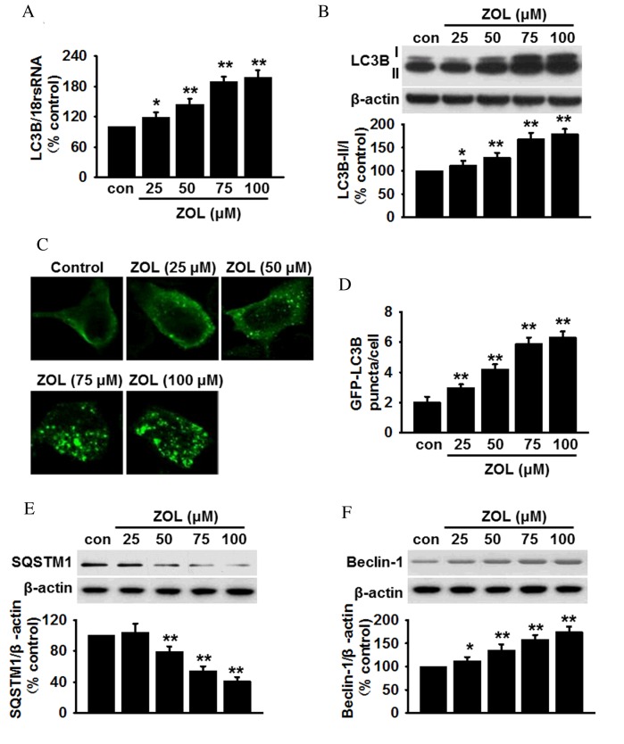
Figure 1.
ZOL induces dose dependent autophagy in HUVECs. (A) Cells were treated with 25, 50, 75 and 100 µM ZOL for 48 h, then the mRNA expression levels of LC3B were determined by reverse transcription-quantitative polymerase chain reaction. (B) The levels of LC3B-I and LC3B-II were analyzed by semi-quantitative western blot. (C) HUVECs were infected with Ad-GFP-LC3 adenovirus prior to ZOL treatment, following which, GFP-LC3 punctae were observed using a laser scanning confocal fluorescent microscope. A representative single cell exhibits LC3 punctae as a marker of autophagic vesicles. (D) Quantification of the mean number of GFP-LC3 punctae per cell. Representative blots and quantitative bar graphs demonstrating the expression of (E) SQSTM1 and (F) Beclin-1, following ZOL treatment. All data are presented as the mean ± standard error. *P<0.05, **P<0.01 vs. untreated control, n=4–6. HUVECs, human umbilical vein endothelial cells; LC3B, microtubule-associated proteins 1A/1B light chain 3B; 18S rRNA, 18S ribosomal RNA; Con, control; ZOL, Zoledronate; SQSTM1, sequestome 1.

