Figure 3.
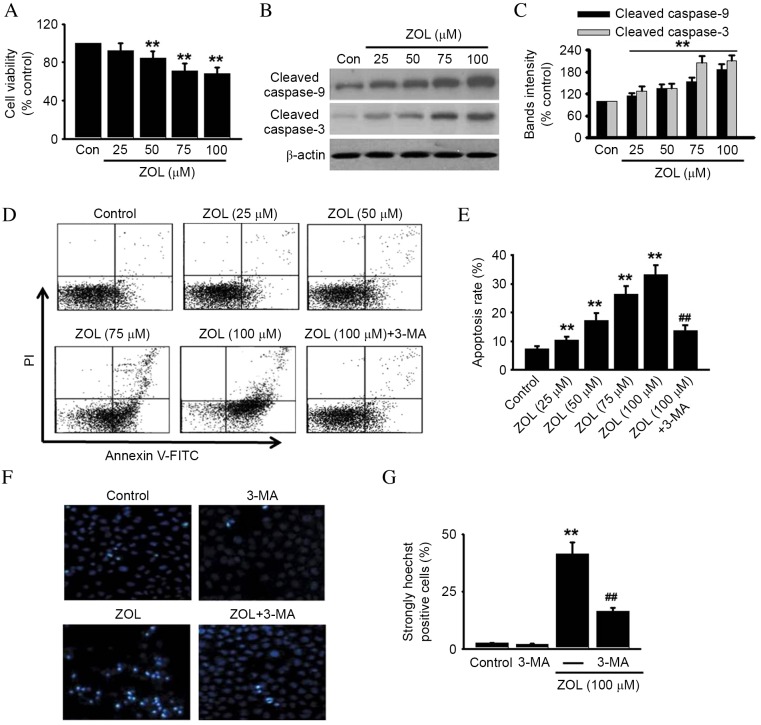
Inhibition of autophagy protects against ZOL-induced HUVEC apoptosis. HUVECs were incubated with various concentrations of ZOL for 48 h, then (A) cell viability was assessed by 3-(4,5-dimethylthiazol-2-yl)-2,5-diphenyltetrazolium bromide (MTT) assay and (B) cleaved caspase-9 and −3 were detected by western blot. Representative western blot images are presented. (C) Densitometric analysis of caspase-9 and cleaved caspase-3 western blots was performed. HUVECs were treated with 5 mM 3-MA prior to ZOL treatment, then (D) the number of apoptotic cells were determined by Annexin V-FITC/PI staining using flow cytometry, (E) followed by quantitative analysis of the percentage of apoptotic cells. HUVECs treated with 5 mM 3-MA and/or 100 µM ZOL were fixed and their DNA stained with Hoechst 33258. (F) Confocal images of nuclear staining are presented and (G) quantification of the number of strongly Hoechst 33258 positive cells was performed. Data are presented as the mean ± standard error, n=6. **P<0.01 vs. Con; ##P<0.01 vs. 100 µM ZOL. HUVECs, human umbilical vein endothelial cells; ZOL, Zoledronate; Con, untreated control; PI, propidium iodide; FITC, fluorescein isothiocyanate; 3-MA, 3-methyladenine.

