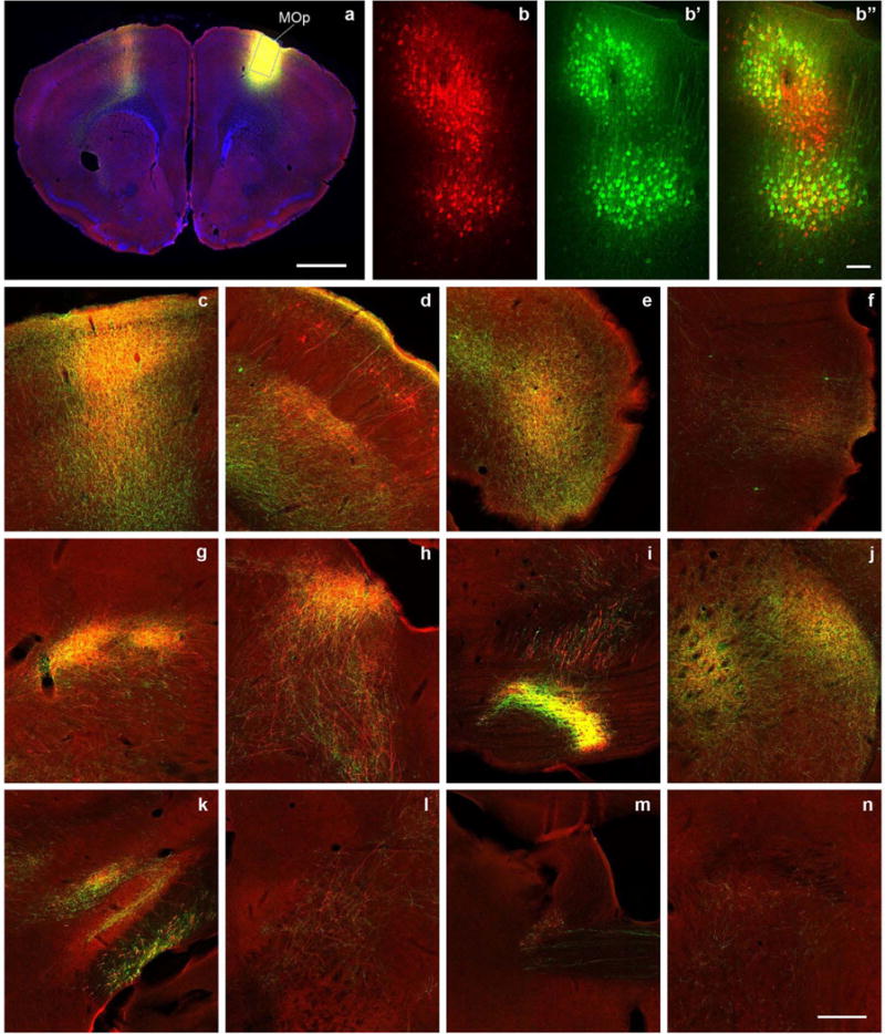
Extended Data Figure 1. AAV/BDA tracer comparison, using a primary motor cortex (MOp) injection as an example.

The cortical and subcortical projections from MOp injection are labelled similarly with the AAV tracer (green) and conventional tracer BDA (red). a, Injection sites of AAV and BDA are mostly overlapping (yellow), with a blue DAPI counterstain. b, Confocal image taken from the box in a shows BDA tracer uptake in individual neurons at the injection site. b’, The same box in a shows AAV infection of individual neurons. b”, Overlay of b and b’ shows the presence of both tracers in the same region and their colocalization in some neurons (yellow). c–f, Examples of cortical projections in the contralateral primary motor cortex, ipsilateral primary somatosensory cortex, agranular insular area (dorsal part), and perirhinal cortex labelled with red, green, or yellow. g–n, Examples of subcortical projections in the ipsilateral ventral posterolateral nucleus of the thalamus and posterior nucleus of the thalamus, superior colliculus, pontine grey, caudate putamen, zona incerta and subthalamic nucleus, midbrain reticular nucleus, parabrachial nucleus, and contralateral bed nucleus of the anterior commissure. Scale bars are 1,000 μm (a); 100 μm in (b, b’ and b”); and 258 μm (c–n). Approximately 18 brain regions were selected throughout the brain to represent broad anatomical areas and diverse cell types (3 cortical and 15 subcortical structures). AAV and BDA were co-injected into each selected brain region in wild-type mice using a sequential injection method developed to target virtually the same anatomical region. For most cases, the anatomical area(s) of tracer uptake are well matched. We found the long-range projections from all studied regions with both tracers. Their patterns were similar between the two tracers in mostly overlapped injection cases. There were more retrogradely labelled neurons with BDA than AAV, although a few retrograde neurons were observed in all studied regions with both tracers. BDA was clearly uptaken by passing fibres in some injections but AAV was not.
