
Figure 5. Topography of cortico-striatal and cortico-thalamic projections.
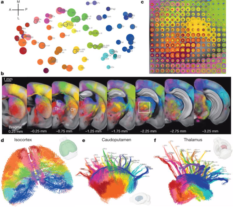
a, Cortical domains in the cortex flat-map. Each circle represents one of 80 cortical injection experiments, whose location is obtained via multidimensional scaling from 3D to allow visualization of all the sites in one 2D plane. The size of the circle is proportional to the injection volume. Clustered groups from Extended Data Fig. 8b are systematically colour-coded. The selected injections for b are marked with a black outline. b, For co-visualization, voxel densities from the 21 selected injections from a are overlaid as ‘dotograms’ at 8 coronal levels for ipsilateral hemisphere. For the dotogram, one circle, whose size is proportional to the projection strength, is drawn for each injection in each voxel; the circles are sorted so that the largest is at the back and the smallest at the front, and are partially offset as a spiral. c, Enlarged view of the dotogram from the area outlined by a white box in b. d, 3D tractography paths in both cortical hemispheres. e, A medial view of 3D tractography paths into the ipsilateral caudoputamen. Voxel starting points are represented as filled circles and injection site end points as open circles. f, A top-down view of 3D tractography paths into the ipsilateral thalamus.