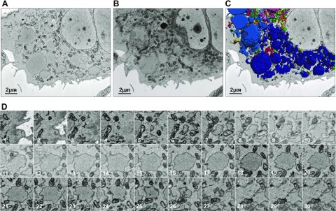
Figure 5.
There is no evidence of interinclusion luminal connectivity by electron microscopy. A, B) Inclusion-laden CHO cell transfected with YFP-Z-α1-antitrypsin and selected using fluorescence microscopy was subjected to serial block-face scanning electron microscopy, single scan (A), and 3-D reconstruction (B). C) Peripheries of Z-α1-antitrypsin-containing inclusions were imaged using Gatan 3View system and combined using Imaris software to generate 3-D projection by isosurface rendering with surface area detail of 36 nm. D) Serial block-face scanning electron microscopy images through inclusion of Z-α1-antitrypsin. Lack of interinclusion luminal connectivity is evident.

