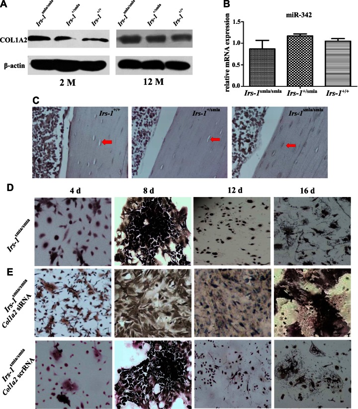
Figure 6.
The relationship of COL1A2 expression in BMSCs, ALP staining in osteocytes, and in the capacity for in vitro osteogenesis in BMSCs derived from Irs-1smla/smla, Irs-1+/smla, and Irs-1+/+ mice. A) Western blots showing that compared to BMSCs from Irs-1+/smla and Irs-1+/+ mice, COL1A2 expression was clearly elevated in BMSCs from Irs-1smla/smla mice at 2 mo but not at 12 mo of age. B) Real-time PCR assays shows that the expression levels of miR-342 showed no significant statistical differences among Irs-1smla/smla, Irs-1+/smla, and Irs-1+/+ mice. C) ALP staining was also increased in osteocytes from Irs-1smla/smla mice compared to osteocytes from Irs-1+/smla and Irs-1+/+ mice. D) BMSCs from the Irs-1smla/smla mice showed low ALP staining after 4 d of osteogenesis induction and then differentiated into osteocyte-like cells after 12 d of osteogenesis induction. These osteocyte-like cells showed a more intense ALP staining than those from WT mice (see Fig. 4E). E) When BMSCs from the Irs-1smla/smla mice were transfected with a Col1a2 siRNA, they lost the ability to differentiate into osteocyte-like cells, even after 16 d of induction.

