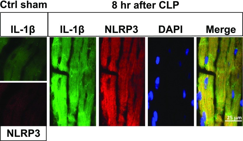
Figure 2.
Activation of NLRP3 inflammasome in LV mouse CMs after CLP. Immunofluorescence analysis of LV frozen sections from sham-procedure cointrol (ctrl sham) mouse WT heart (left boxes) or hearts 8 h after CLP. Magnification, ×63. Analysis involved immunostaining for IL-1β (green), NLRP3 (red), nuclear staining by DAPI, and the merge image for all 3 stains. Staining for IL-1β and NLRP3 was very faint in sham-procedure control hearts, in striking contrast to hearts 8 h after CLP.

