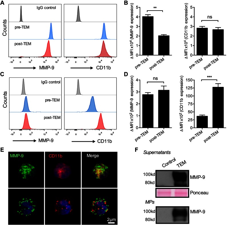
Figure 2.
MMP-9 localizes to both PMN granules and cell surface and is secreted during PMN TEM. Expression of MMP-9 on PMNs was examined by using flow cytometric analysis. A, B) PMNs before and after TEM were fixed, permeabilized, and stained for MMP-9 and CD11b. Representative flow diagrams (A) and quantification (B) show a significant decrease in total levels of MMP-9 in PMNs after TEM. **P < 0.01. C, D) Analysis of surface expression of PMNs before and after PMN TEM (under nonpermeabilized conditions) revealed detectable basal levels of surface MMP-9. PMN surface expression of MMP-9 was not significantly altered during TEM. ns, not significant. ***P < 0.001. E) Representative immunofluorescence images show total (top) and surface MMP-9 (bottom) expression by permeabilized and nonpermeabilized PMNs, respectively. MMP-9 (green) on PMN surface did not colocalize with CD11b (red). F) Release of MMP-9 by transmigrating PMNs was further confirmed by Western blot analysis. Secreted MMP-9 was detected in cell supernatants (soluble form) and associated with PMN-MPs after PMN TEM. Equal protein loading in cell supernatants was confirmed with Ponceau protein stain. For all panels, n = 3 independent experiments.

