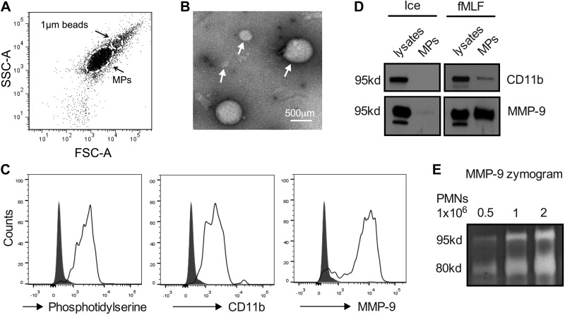
Figure 3.
Activated PMNs secrete microparticles with enzymatically active MMP-9. MPs were isolated from supernatants of fMLF-stimulated PMNs (1 μM, 20 min, 37°C). A) The presence of PMN-derived MPs (<1 μm) in supernatants of activated PMNs was confirmed by flow cytometry. SSC, side scatter; FSC, forward scatter. B) Transmission electron microscopy micrograph depicts the heterogeneity in the size of PMN-derived MPs. C) PMN-MPs were further characterized by flow cytometry for the expression of membrane marker, annexin V and the specific myeloid marker CD11b. PMN-MPs that stained positive for both annexin V and CD11b were also found to express high levels of MMP-9. D) CD11b and MMP-9 expression on MPs derived from fMLF-stimulated PMNs was also confirmed by Western blot analysis. E) Gel zymography confirmed that MP-bound MMP-9 was enzymatically active. MMP-9 enzymatic activity (as evident from collagen degradation) was enhanced with increasing concentration of MPs. All panels are representative of at least 3 independent experiments. Ice, unstimulated PMNs kept on ice at all times.

