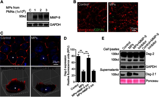
Figure 4.
PMN-MP binding to IECs leads to MMP-9-dependent cleavage of Dsg-2. To examine the effect of PMN-MP binding to IEC membrane on Dsg-2 expression and cleavage, PMN-MPs were added to both the upper and lower chambers of cell-migration assay plates, so that both the basolateral and apical IEC surfaces were exposed to treatment (3 h, 37°C). A) MPs derived from an increasing number of PMNs (as indicated) were incubated with IECs, and their binding was examined by Western blot analysis. B) PMN-MP (derived from 2 × 10⁶ PMNs) binding to IECs was further examined with fluorescence microscopy. Representative immunofluorescence images show that PMN-MPs (positive for CD11b, green) bound to IECs with high efficiency. The IEC surface was visualized with a membrane stain, cell mask (orange). C) Representative immunofluorescence images (top) show that PMN-MP binding to epithelial cells led to decreased expression of Dsg-2 (red: Dsg-2; blue: nuclei). A Z-stack 3-dimensional surface plot (bottom) shows decreased levels of Dsg-2 expression at the basolateral/apical cell borders. D) Quantification of the relative Dsg-2 expression based on fluorescence staining from 3 independent experiments (n = 50 cell/condition). **P < 0.01. E) Western blot analysis confirmed that PMN-MP binding to IECs induced cleavage of Dsg-2. Although constitutive low-level cleavage of Dsg-2 by IECs was observed, treatment with PMN-MPs dramatically increased the abundance of a 100-kDa Dsg-2 cleavage product (Dsg-2 f) both in cell supernatants and in cell lysates. PMN-MP-mediated cleavage of Dsg-2 was MMP-9 dependent, as it was reversed in the presence of an MMP-9 inhibitor (MMP-9 inhibitor II, 100 nM). Constitutive IEC-mediated cleavage of Dsg-2 was similarly suppressed in the presence of MMP-9 inhibitor. Blots are representative of 4 independent experiments.

