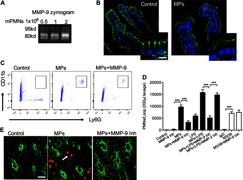
Figure 7.
PMN-MP treatment of murine intestines promotes PMN TEM in vivo. Murine MPs were prepared from bone marrow–derived PMNs. A) Gel zymography was used to confirm that mPMN-MPs expressed enzymatically active MMP-9. MMP-9 enzymatic activity (as evident from collagen degradation) was enhanced with an increasing concentration of MPs. B) To examine the effect of mPMN-MPs on Dsg-2 expression and distribution in vivo, mPMN-MPs were administered intraluminally into ligated ileal loops (MPs were derived from 5 × 10⁶ PMNs for 3 h). After PMN-MP incubation, tissue from ileal loops was prepared for immunofluorescence imaging analysis. A dramatic loss of Dsg-2 (green) from the apical junctional regions was observed. Scale bar, 50 µm. C–E) In separate experiments, after mPMN-MP treatment, PMNs that migrated into the lumen were isolated by lavage and quantified using flow cytometric analysis. C) Representative flow diagrams (n = 5 mice/condition) depict mPMN-MP-induced PMN migration into the intestinal luminal space. PMN migration was further potentiated with the addition of LPS or M3/38-mediated down-regulation of Dsg-2. D) Quantification of data from C. E) Representative immunofluorescence images of cryosections from the ileum show PMN infiltration (red) of the intestinal epithelia (outlined by actin staining, green) and the lumen after mPMN-MP treatment (n = 5 mice/condition). Scale bar, 50 µm.

