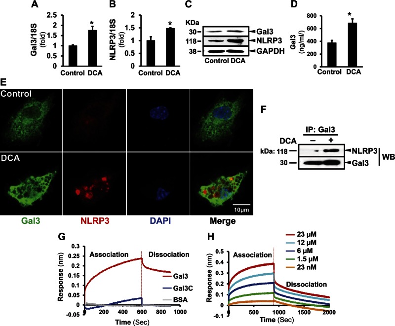
Figure 3.
DCA induces Gal3 and NLRP3 expression and their association in hepatic macrophages. Primary macrophages were isolated from mouse livers and treated with DCA (100 µM for 24 h). A, B) RT-qPCR showed significantly induced Gal3 (A) and NLRP3 (B) expression after DCA treatment (means ± sem; n = 4). *P < 0.05. C) The induction at protein levels was also detected by Western blot analysis. D) Culture medium was collected for ELISA to examine Gal3. The DCA-challenged cells released significantly higher levels of Gal3 (means ± sem; n = 6). *P < 0.05. E) To visualize Gal3 and NLRP3 in macrophages, immunofluorescence studies were done in the DCA-treated macrophages. The signals for Gal3 (green) and NLRP3 (red) were dispersed and weak in nontreated cells. In the DCA-challenged cells, enhanced Gal3 and NLRP3 were seen, and more NLRP3 clusters were observed. F) Immunoprecipitation of Gal3 and NLRP3 Western blot showed their association in DCA-treated macrophages. BLI was conducted to confirm the association. Recombinant NLRP3 loaded sensors were incubated with either recombinant WT Gal3 or N-terminal truncated Gal3 (Gal3-C) (18 μM). The association was recorded for 600 s. G) The sensors were moved to the blank buffer to measure the dissociation. Gal3 showed an obvious association/dissociation pattern. This was not present in Gal3-C. Thus, Gal3 directly interacts with NLRP3, and its N-terminal motif is crucial for this interaction. H) When the NLRP3-loaded sensors were incubated with Gal3 at a series of concentrations, a dose-dependent association was observed.

